A Surgical Camera that Keeps an Eye Out for Tumours

A group of German researchers from the Fraunhofer Institute for Manufacturing Engineering and Automation have developed a new camera for differentiating cancerous tissue from healthy tissue in the body. The new camera will come in handy to surgeons operating on cancer patients to recognize and remove the entire tumour without leaving behind tiny cells which they may otherwise miss.
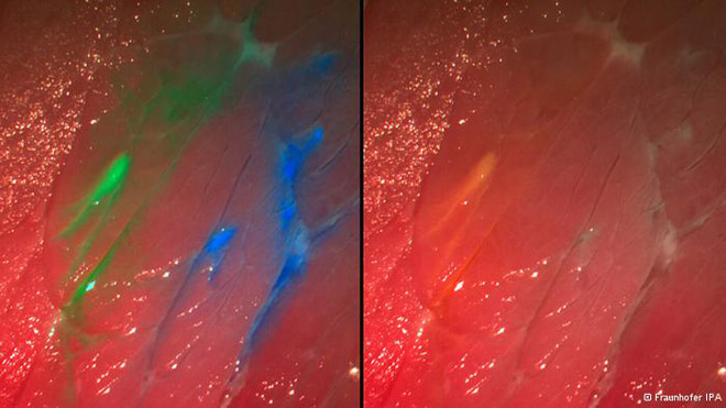
In order to use this new surgical camera, first the patient has to be injected with dyes- fluorescent molecules carrying antibodies that attaches itself to tumour cells. These dyes glow in different colours when exposed to light of specific wavelengths. During the operation, light of wavelength corresponding to the dye is used to illuminate the tissue. The affected tissue with fluorescent molecules attached to it starts glowing; the camera captures and superimposes this image with a normal colour image, to provide accurate information about the tumour cells or metastases. This helps surgeons to completely remove only the effected portions, leaving healthy tissue behind.
The new surgical camera is capable of handling up to 4 different dyes at a time, a feature that helps in studying the biochemical structure of tumour by observing its interaction with multiple dyes. The camera is currently in a prototype stage, and it will soon be available for integration with surgical microscopes and endoscopes.
Source: Fraunhofer IPA